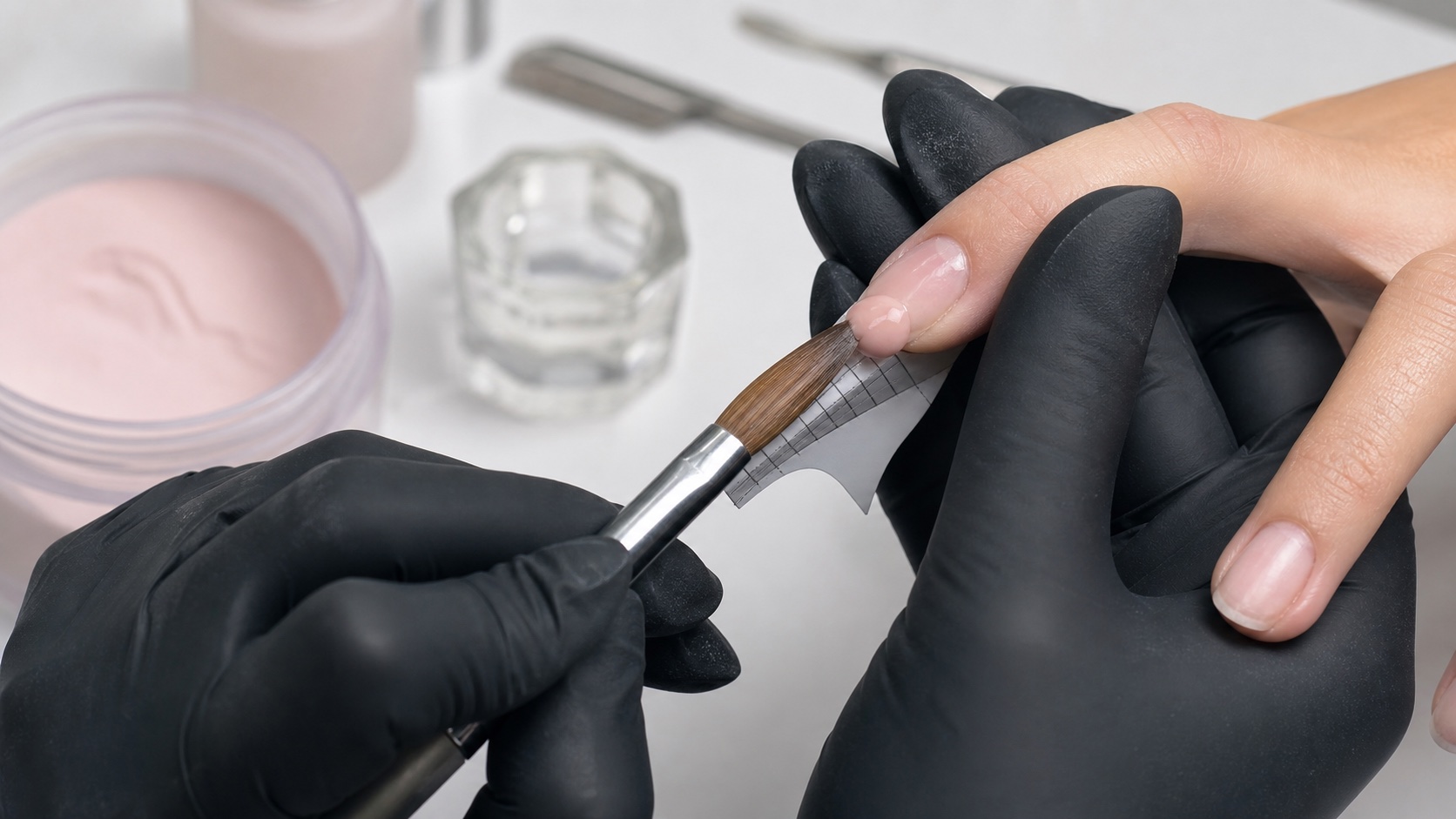
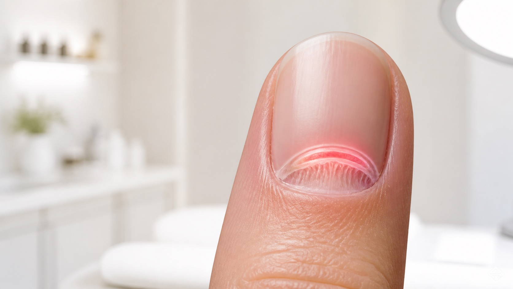
The nail matrix is living tissue that produces most of the nail plate—the hard, translucent structure commonly called the nail. Most of the matrix is hidden beneath the proximal nail fold, while its distal portion may be visible as the lunula. Because new plate is formed at the base and moves forward gradually, an event affecting the matrix can become visible days or weeks later and then travel towards the free edge as the nail grows.
Quick answer: the matrix makes the nail plate; the nail bed supports the plate that has already formed; the proximal fold and cuticle help protect the growing area. A nail professional can observe the external nail unit and document change, but cannot diagnose matrix disease or confirm the cause of a ridge, colour change or deformity.
This anatomy guide opens the BeautyLearn professional nail guide library. It owns the broad nail-matrix intent and does not replace the more specific guides that will cover cuticle anatomy, nail preparation, visible nail changes and service technique. To build anatomy and practical skills in sequence, compare the English-market online nail courses or review the curriculum and current availability of the complete nail technician course. English courses and instructors are separate records and may differ from the Italian catalogue.
What the nail matrix is
The nail matrix is specialised germinative epithelium. Its cells proliferate, keratinise, flatten and become tightly packed onychocytes. These cells form the nail plate and are pushed distally as newer material is produced behind them. The matrix therefore belongs to the living nail unit, whereas the completed plate itself is keratinised material without living cells in the everyday sense.
The matrix is not the whole pale area at the base of the nail, and it cannot be inspected directly during an ordinary manicure. The NCBI nail histology review describes it as the origin of the nail plate. Its proximal portion contributes most of the plate, while the distal portion contributes deeper layers. This is why injury or surgery in the matrix carries a risk of persistent nail-plate change.
Nail anatomy varies normally among people and among fingers. Lunulae may be obvious on the thumbs and barely visible elsewhere. Plate width, curvature, thickness, surface ridging and growth speed also vary. A small or invisible lunula alone is not proof that the matrix is weak, blocked or unhealthy.
Matrix, lunula, nail bed, plate and cuticle: the differences
One nail unit, different structures
Production, protection and support are not interchangeable
Matrix
Living plate-producing tissue
Lunula
Sometimes-visible distal matrix
Nail plate
Compact keratinised structure
Nail bed
Support beneath the plate
Fold + cuticle
Protective proximal seal
Structure | Main role | Professional implication |
|---|---|---|
Nail matrix | Produces most of the nail plate | Mostly hidden; do not claim to inspect or stimulate it directly |
Lunula | Visible distal portion of the matrix in some nails | Visibility varies and is not a stand-alone health test |
Nail bed | Supports and anchors the plate as it advances | Do not probe beneath an attached plate or cover unexplained separation |
Nail plate | Protective keratinised structure visible at the surface | Preparation removes surface contamination and shine, not layers indiscriminately |
Proximal nail fold | Living skin that covers and protects the proximal matrix | Pain, cuts and aggressive abrasion are not acceptable preparation |
Cuticle | Keratinised seal that adheres to the emerging plate | Distinguish removable non-living residue from living tissue |
The NCBI overview of nail anatomy explains how the proximal fold protects the matrix and how the nail bed extends from the lunula towards the hyponychium. Terminology is important because a technician who calls every proximal structure ‘cuticle’ may remove living tissue or give the client a misleading explanation of what was treated.
How the nail grows from the matrix
The plate advances from the base
A visible change may reflect an earlier event
Formation
Matrix cells produce keratin
Hidden phase
New plate remains under the fold
Emergence
A change appears near the base
Advance
The mark travels with growth
The plate does not grow from its free edge. New keratin is formed at the matrix, and the plate advances over the nail bed. Fingernails grow at roughly 3 millimetres per month on average, while toenails are slower at around 1 millimetre per month. These figures describe population averages, not a deadline for one client. Digit, age, circulation, illness, medication, season, trauma and individual biology can all affect the rate.
A fingernail may need about six months to replace its visible length; a toenail can require twelve to eighteen months. This explains why a proximal mark can remain visible for a long time even after the initiating event has ended. It also explains why cosmetic promises to ‘repair the matrix in days’ or make a damaged nail grow out immediately are not credible.
Growth should be documented by position, date and a consistent photograph when monitoring is appropriate. A mark that moves towards the free edge with the plate tells a different story from pigmentation or disruption that remains fixed at the base or continues to widen. Movement can provide useful information to a clinician, but it does not establish a diagnosis in a salon.
Why matrix changes may appear later
The part of the plate being formed is hidden beneath the proximal fold. If matrix production is temporarily interrupted, the affected keratin must first emerge from under that fold before the change can be seen. A person may therefore connect a new groove or irregularity with yesterday’s manicure even when its position suggests that the event occurred weeks earlier—or may blame an older event that is unrelated. Timing is evidence to examine, not proof of cause.
A broad temporary interruption may eventually appear as a transverse depression; a more severe interruption can be associated with partial shedding. Local trauma may produce a focal ridge, split or colour change. Similar-looking signs can also have dermatological, systemic, infectious or medication-related causes. The nail professional should describe location, number of nails, onset, symptoms and change over time without converting those observations into a medical label.
What can damage or irritate the protected area
The matrix is protected, but the proximal unit is not immune to trauma. Crushing injuries, repeated picking, forceful pushing, cuts, burns, severe inflammation and inappropriate instrument use can affect the tissues around it. In a salon, preventable risks include driving a tool beneath the proximal fold, using excessive pressure or speed with an e-file, repeatedly abrading the same spot, forcing adhered tissue and continuing while the client reports sharp pain or heat.
A clean-looking proximal pocket is not a valid reason to invade living tissue. Product placed too close to or onto the skin can also cause irritation and increases exposure to uncured material. Technique should maintain visibility, stable support, low pressure and a clear distinction between plate surface, non-living residue and living fold.
Nail cosmetics do not enter the hidden matrix to feed it. Oils and moisturisers can support the surrounding skin and improve surface flexibility; they cannot regenerate scarred matrix tissue or treat an underlying disorder. Avoid language such as ‘activates the matrix’, ‘detoxifies the nail’ or ‘heals from within’ unless a specific, lawful product claim is supported by appropriate evidence.
Professional assessment before a nail service
Observe before tools and product
External assessment leads to a service decision
Light and bare-nail view
Compare digits and both hands
Ask about onset and symptoms
Record facts with consent
Proceed, adapt, postpone or refer
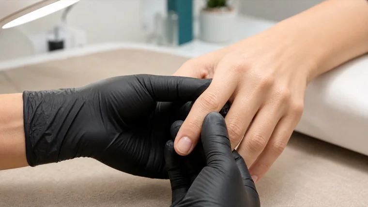
Assessment is external, non-invasive and linked to a decision: proceed, adapt, postpone or refer. Remove existing product only when it can be done safely and when seeing the underlying plate is necessary. Use good light and compare both hands. Ask about recent trauma, pain, bleeding, sensitivity, changes under the coating, previous reactions and whether a clinician is already involved.
Observe the plate, proximal and lateral folds, surrounding skin and free edge before using tools.
Compare all ten nails and note whether the change affects one digit, several digits or the regrowth itself.
Ask when it began, whether it is changing and whether pain, swelling, discharge, bleeding or loss of function is present.
Record only relevant facts with consent: date, site, client report, photograph and the decision taken.
Do not cover a sign merely to make it invisible. If the cause or safety is unclear, postpone and recommend appropriate medical assessment.
When not to cover the nail
Visibility can be safer than camouflage
Stop criteria protect the client and the evidence
Proceed
Intact, comfortable, understood
Adapt
Minor cosmetic variation within scope
Postpone
Unclear change or compromised skin
Refer
Pain, injury or concerning evolution
Postpone a cosmetic service when there is active bleeding, an open wound, significant pain, marked swelling, pus or discharge, sudden lifting after trauma, a displaced plate, a rapidly changing dark mark, unexplained distortion emerging from the base or any presentation outside the technician’s competence. The purpose is not to identify the condition; it is to avoid obscuring it, aggravating it or delaying care.
After an acute injury, the American Academy of Dermatology advises medical attention for severe pain, inability to bend the digit, extensive blood beneath the nail or a markedly dark injury. Local emergency guidance and the client’s clinician remain the appropriate source for individual care. A technician should not drain blood, prescribe medication, remove a traumatised plate or attempt to ‘release pressure’.
Once a clinician has assessed the area, proceed only within the written or clearly understood limits relevant to the service, and only if the skin is intact and the client is comfortable. A medical green light does not remove the need for gentle technique, product compatibility and continuing observation.
Common myths about the nail matrix
Myth | More accurate interpretation |
|---|---|
The visible lunula is the whole matrix | Most of the matrix is hidden beneath the proximal fold |
No lunula means poor health | Lunula visibility varies normally and cannot diagnose health |
A ridge proves the matrix was damaged | Ridges have multiple possible causes; timing and clinical assessment matter |
Gel stops the nail from breathing | The plate does not breathe; safe service depends on assessment, chemistry, curing and removal |
Oil makes the matrix grow faster | Oil conditions exposed skin and plate; it does not command matrix cell division |
Nail-matrix checklist for technicians
Use clear, even lighting and examine the bare nail whenever safety depends on it.
Distinguish plate, living fold and removable keratinised residue before choosing a tool.
Keep pressure, speed and depth controlled; pain is information, not a hurdle.
Describe visible facts and changes over time without naming a disease.
Photograph only with consent, protect the record and avoid diagnostic captions.
Postpone and refer when pain, injury, inflammation or unexplained change makes cosmetic work inappropriate.
Frequently asked questions about the nail matrix
Where is the nail matrix located?
Most of it sits beneath the proximal nail fold at the base of the nail. Only the distal portion may be visible through the plate as the lunula.
Is the lunula the same as the matrix?
The lunula is the visible distal part of the matrix, not the whole structure. A large part remains protected under the proximal fold.
Does every healthy nail have a visible lunula?
No. Lunula size and visibility vary by person and digit. Its absence from view does not diagnose a deficiency or matrix problem.
How quickly do nails grow?
Fingernails average around 3 millimetres per month and toenails around 1 millimetre, but individual rate varies. Full replacement takes months, not days.
Can a damaged nail matrix recover?
Outcome depends on the cause, site, depth and duration of injury. A temporary insult may grow out; scarring can produce lasting change. Only an appropriately qualified clinician can assess an injury and prognosis.
How can I tell whether the matrix is damaged?
You cannot confirm it from one surface sign. Recurrent change beginning at the base, significant trauma, pain or persistent distortion warrants medical assessment rather than a salon diagnosis.
Can manicure tools reach the matrix?
The matrix is protected beneath the fold, but forceful or invasive work around the proximal area can injure protective tissues and may affect deeper structures. Tools should never be driven under living skin.
Does removing the cuticle damage the matrix?
Careful removal of detached non-living residue is different from cutting or abrading living proximal tissue. Aggressive work can break the protective seal and cause injury or inflammation.
Can nail oil stimulate the matrix?
Oil can condition exposed skin and improve the feel of the plate and surrounding tissues. It cannot directly control hidden matrix growth or repair a medical disorder.
Why can a groove appear weeks after an event?
The affected plate is formed under the proximal fold and must move forward before it becomes visible. The location of a groove may help estimate timing, but does not prove the cause.
Should a ridge or colour change be covered with gel?
Not automatically. First assess whether the nail is intact, comfortable and within cosmetic scope. New, painful, dark, widening or unexplained change should remain visible and be assessed medically.
Can a nail technician diagnose matrix disease?
No. A technician can observe, document, postpone a service and recommend medical assessment. Diagnosis and treatment belong to qualified healthcare professionals.
What should be recorded before postponing a service?
Record only relevant visible facts, the client’s report, date, affected digits, symptoms and the decision. Photographs require consent and secure handling; avoid speculative diagnostic language.
When does a nail injury need urgent care?
Severe pain, extensive blood beneath a nail, an open or displaced nail, inability to move the digit or significant swelling requires prompt assessment under local medical guidance.
Last reviewed: 5 September 2026. Educational content based on current anatomical and dermatological sources. It does not diagnose nail conditions, determine the cause of visible change or replace individual medical advice.
Next technique: use the professional nail-filing guide to connect anatomy with grit choice, a controlled sequence and three-view shape checks.
For repeated biting or picking, use the separate nail-biting and onychophagia guide, which explains triggers, tissue effects, behaviour-change evidence and professional stop criteria without diagnosing.