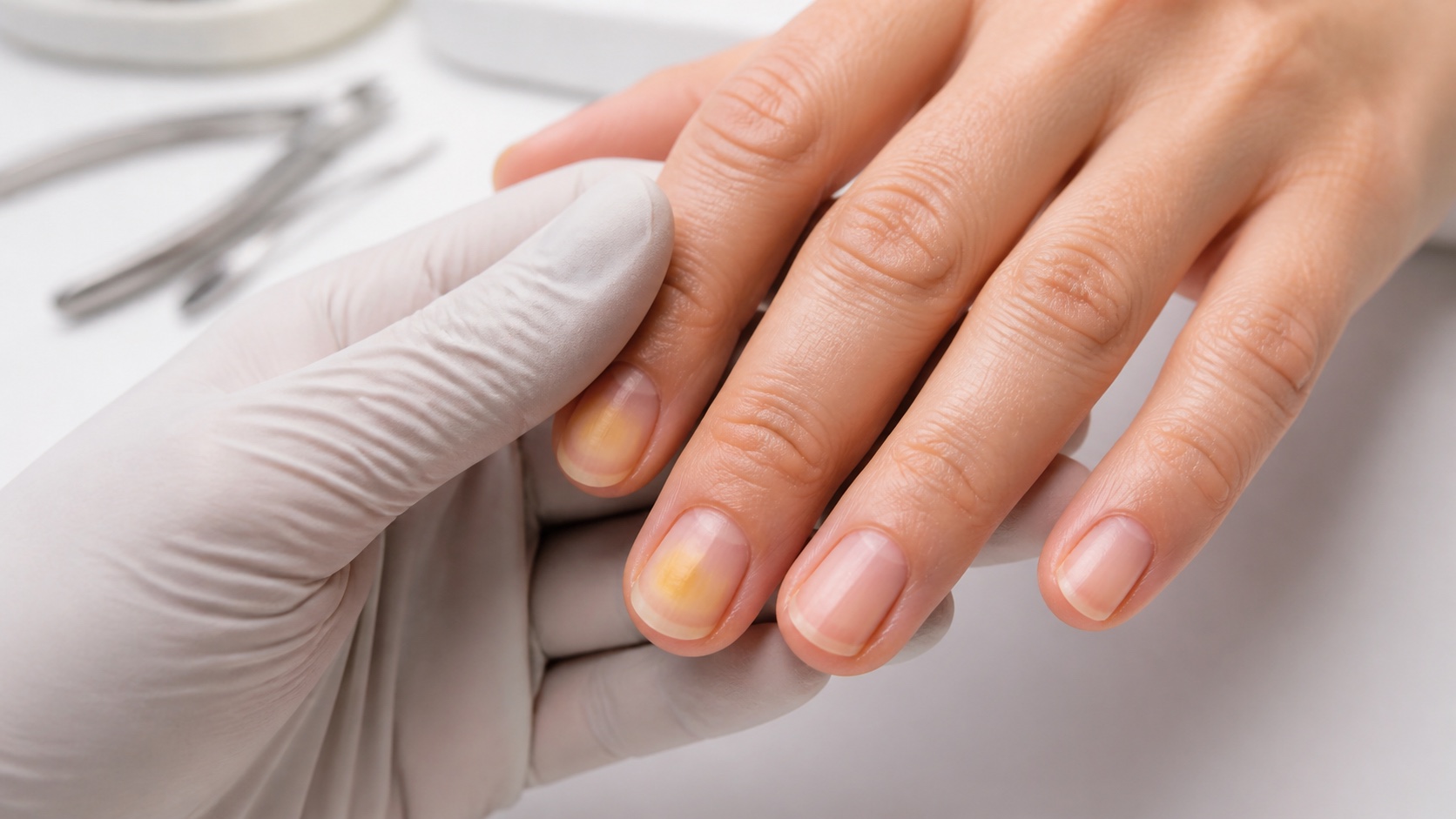
Yellow nails can reflect a superficial stain, a change within the nail plate or material beneath a lifted nail. Colour alone cannot identify the cause. Distribution, surface, thickness, growth, separation, discomfort, surrounding skin, product history and change over time all matter. Treating every yellow nail as fungus—or filing until the colour disappears—can damage the plate and delay the right assessment.
Quick answer: an even stain after strongly pigmented polish, on an otherwise smooth nail, may grow toward the free edge with the plate. Yellowing with thickening, crumbling, lifting, debris, pain or inflamed skin needs healthcare assessment rather than gel, polish or an extension. Green or green-yellow colour under a lifted area should not be called “mould” or covered. A photograph cannot confirm fungus, psoriasis or another disease.
This guide helps clients, learners and nail professionals describe changes and choose a safe next step; it does not diagnose or recommend treatment. Read the nail-matrix anatomy guide for growth structures, the nail-lifting guide for product adhesion and the BeautyLearn professional nail guide library for adjacent intents. English courses and teachers remain separate market records.
Yellow is an observation, not a diagnosis
Locate the colour before naming a cause
Surface, plate and sub-nail colour mean different questions
Surface
Pigment or external exposure
Plate
Texture, thickness and growth
Under plate
Lifting, debris or bed colour
Context
Products, trauma and symptoms
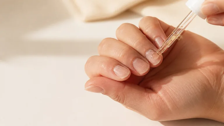
First locate the colour. It may sit on the surface after polish, pigment, nicotine, dye or occupational exposure; appear within a changed plate; or show through from the nail bed or material beneath a separated edge. Then compare all nails, hands and feet. One recently polished fingernail tells a different story from several slowly growing, thickened toenails.
Use neutral daylight or a high-quality neutral task light and remove removable colour gently. Record when the change began, which products were worn, removal method, trauma, water exposure, footwear, pain, medicines and known health conditions only as the person reports them. Do not scrape underneath, aggressively buff the surface or use a colour chart to name a disease.
Pattern | What it may suggest | Safe response |
|---|---|---|
Smooth, even stain after dark polish | Possible superficial pigment transfer | Photograph, protect and observe growth |
Yellow-white, thick, brittle or crumbly nail | Fungal infection is one possibility among others | Clinical assessment and testing where indicated |
Pits, lifting, yellow-brown colour or sub-nail build-up | Psoriasis or another inflammatory process may be considered | Dermatology assessment |
Many thick, slow-growing yellow nails plus swelling or respiratory symptoms | Rare systemic pattern needs medical review | Medical assessment; urgent care for severe breathing symptoms |
Surface staining from polish and external pigments
A plausible stain should move with growth
Observe rather than sand away the evidence
Confirm recent pigment exposure
Check surface and thickness
Photograph in neutral light
Observe growth without deep buffing
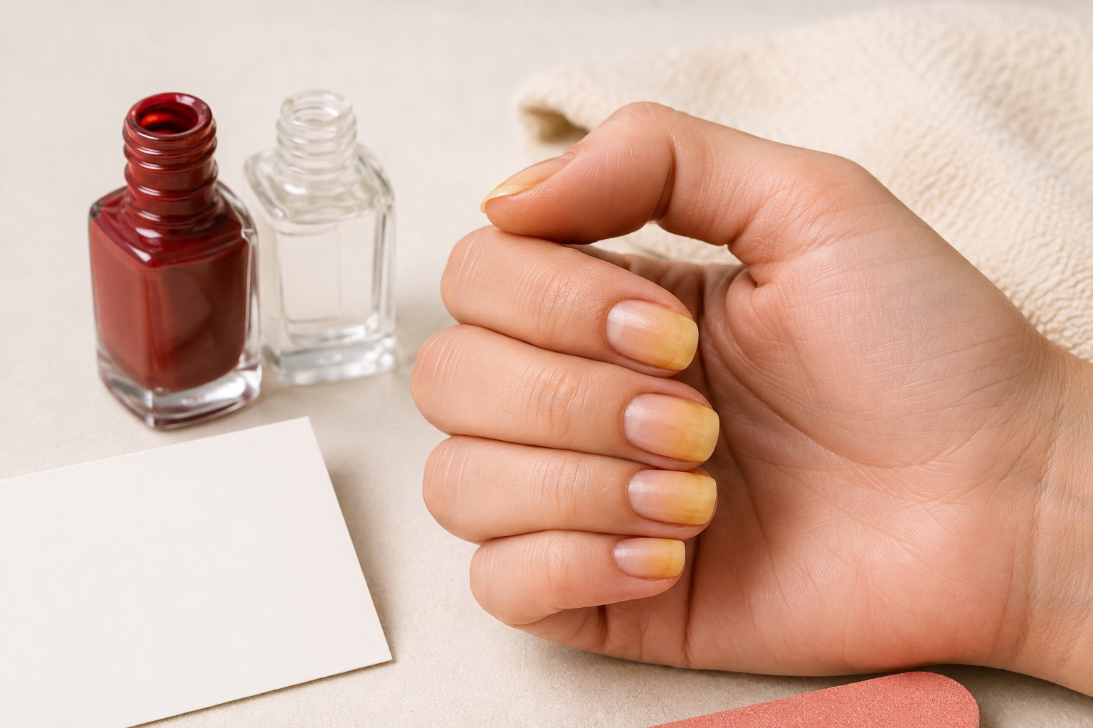
Strong red, orange, brown and dark pigments can leave a yellow or orange cast after repeated wear, especially when the system and base have not prevented migration. Nicotine, hair dyes, art pigments, cleaning substances and occupational colour can also affect exposed fingers. A cosmetic stain is more plausible when the plate stays smooth and normally thick, surrounding skin is comfortable and the discoloured section advances with growth.
“More plausible” is not a diagnosis. Remove polish with the correct product, wash and dry, photograph all nails and compare at sensible intervals. Do not bleach the plate or grind away colour. Aggressive surface filing creates new thinning, white patches and sensitivity that make later observation harder. A clear base may reduce future pigment transfer but cannot guarantee prevention across every colour and nail.
Fungal nail infection: appearance is not confirmation
Onychomycosis can make a nail white, yellow, orange or brown and may add thickening, crumbling, distortion, debris or separation. It more often affects toenails, but colour and texture overlap with trauma, psoriasis and other disorders. The UK NHS describes brittle, discoloured or thickened nails as possible symptoms; it does not imply that every such nail has fungus.
The American Academy of Dermatology explains that dermatologists may clip, scrape or sample material for microscopic examination because psoriasis or injury can resemble fungal disease. Treatment choice depends on the person, extent, organism and health context. A nail professional should not recommend an antifungal from a photograph, share tools between affected and unaffected nails or cover a suspicious change while waiting to see whether product “holds”.
Use the current NHS fungal nail overview for patient-facing signs and the AAD’s diagnosis and treatment explanation for why clinical testing may be necessary. Do not copy prescription, duration or home-treatment advice into a cosmetic consultation.
Nail psoriasis and inflammatory changes
Nail psoriasis may occur with or without obvious skin plaques. The AAD lists pits, white-yellow-brown discolouration, crumbling, lifting, sub-nail build-up and blood beneath the plate among possible signs. None is exclusive to psoriasis. Joint pain, swelling or morning stiffness in a person with possible skin or nail psoriasis belongs in a medical history, not a salon conclusion.
Refer persistent or changing patterns for dermatology review and avoid deep preparation over an altered plate. The AAD’s current nail psoriasis guidance emphasises slow nail improvement and tailored treatment. A cosmetic overlay cannot establish whether a change is stable, infectious, inflammatory or caused by trauma.
Lifting, trauma and colour beneath the plate
Onycholysis means separation of the nail plate from the bed. The separated portion often looks opaque white but may appear yellow or pinkish, and keratin or debris can accumulate beneath it. Repeated impact, long levers, aggressive preparation, sharp cleaning beneath the nail, chemical irritation, skin conditions and infection are among possible contexts. Do not probe, reattach or seal the pocket.
A green, green-yellow or dark green area beneath a lifted nail is often associated clinically with Pseudomonas pigment. It should not be labelled “mould”, buffed until pale or sealed under enhancement. Moisture trapped between plate and product can support microbial growth. A dedicated onycholysis and green-nail guide will own that detailed intent; here the rule is stop, document, keep the area uncovered and refer according to symptoms and local scope.
Yellow nail syndrome is rare
Yellow nail syndrome is not a synonym for ordinary yellow nails. A recent PubMed-indexed clinical overview describes it as a rare disorder associated with yellow nail changes, lymphoedema and respiratory tract disease; not all features necessarily appear together. An established review describes slow-growing, thickened, curved nails, loss of cuticle and possible separation, often affecting multiple nails.
Do not raise this rare condition because one nail is stained. Many nails that grow unusually slowly, especially alongside limb swelling, chronic cough or other respiratory symptoms, warrant medical assessment. Severe shortness of breath or acute symptoms require urgent local care. The yellow nail syndrome review provides clinical context but cannot be used by a salon to screen or diagnose.
Warning signs that change the next step
Structure and symptoms outweigh colour
Do not cover a changing nail
Structure
Thick, brittle, crumbly or detached
Inflammation
Pain, heat, swelling or discharge
Distribution
Several nails or progression
Systemic
Swelling or respiratory symptoms
Colour persists or progresses without a clear external pigment history.
The nail becomes thicker, brittle, crumbly, distorted or detached.
Pain, heat, swelling, discharge, odour or spreading redness is present.
A new or changing dark streak appears, especially with pigment on surrounding skin.
Several nails change together or growth becomes unusually slow.
The person has diabetes, reduced circulation or immune suppression, or reports relevant medication or illness.
The AAD’s nail changes a dermatologist should examine includes lifting, redness and swelling, and new or changing dark streaks. Intense pain, fever, rapidly spreading redness, red streaking or acute breathing difficulty require urgent assessment through the local healthcare route.
Professional observation and referral protocol
Describe what you see; do not diagnose
A safe pause preserves the evidence
Observe all nails in neutral light
Record pattern and reported history
Apply stop criteria
Refer and keep the change visible
Remove removable colour gently only when safe and authorised; do not file to reveal a presumed diagnosis.
Observe all nails and surrounding skin in neutral light, comparing hands and feet only within scope.
Ask when it began, what products or exposures preceded it and whether pain, swelling, odour, discharge or health changes are present.
Describe location, colour, surface, thickness, separation and reported symptoms without writing “fungus”, “psoriasis” or another diagnosis.
Postpone polish, gel, acrylic, adhesives and aggressive preparation when the change is unexplained or stop signs are present.
Recommend the appropriate healthcare route, record the decision and protect any photographs as health-related personal data.
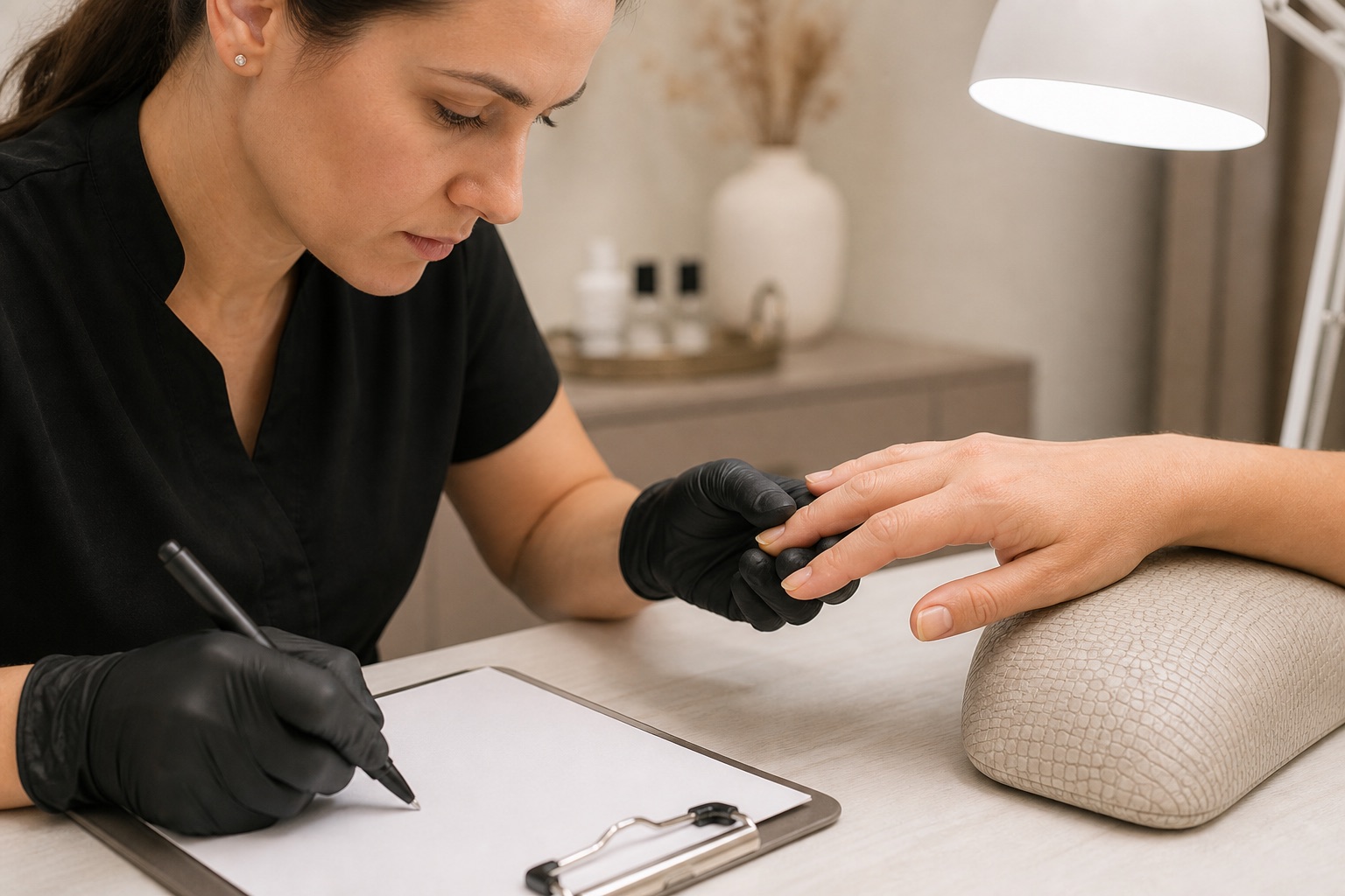
Professional stop-or-proceed matrix
Presentation | Decision | Reason |
|---|---|---|
Known polish stain; smooth, intact, comfortable plate | Conservative service may be considered after assessment | Pattern and history are consistent, but still document |
Unexplained persistent yellowing | Postpone and recommend clinical review | Colour alone cannot establish the cause |
Thickening, crumbling, debris or separation | No cosmetic cover; refer | Fungus, psoriasis, trauma and other causes overlap |
Pain, discharge, inflammation or green colour | Stop and seek prompt healthcare advice | Possible infection or injury lies outside cosmetic scope |
What to do safely while waiting for assessment
Leave an unexplained change visible, keep the nail reasonably short without entering attached tissue, keep hands or feet clean and dry, and avoid sharing files or tools. Use gloves appropriate to wet work without leaving damp hands enclosed for long periods. Do not pull detached plate, dig beneath it or repeatedly photograph it under different colour casts.
Avoid bleach, concentrated acids, harsh whitening mixtures, deep buffing and unverified essential-oil or supplement claims. Ask a pharmacist or clinician before using antifungal medicine: correct treatment depends on diagnosis, site, extent, other conditions and medicines. A product that changed the colour is not proof that the underlying cause was treated.
Frequently asked questions about yellow nails
Why do nails turn yellow after polish?
Strong pigments can stain the surface, especially after repeated wear. A smooth stain may grow out, but persistent or structurally changed nails need assessment.
Are all yellow nails fungal?
No. Pigment, trauma, psoriasis, lifting and other conditions can look similar. Fungus may require a clinician’s examination and sample.
Can I diagnose nail fungus from a photo?
No. Lighting and image processing distort colour, and several disorders overlap. A clinician may need microscopy or other testing.
Should I buff away yellow staining?
Do not file deeply to chase colour. It can thin the plate and obscure later observation. Use gentle surface care only when the change is clearly understood.
Can a base coat prevent yellow nails?
A compatible base may reduce pigment transfer, but cannot guarantee prevention and does not protect against non-cosmetic causes.
Can I cover a yellow nail with gel polish?
Not when colour is unexplained or accompanies thickening, lifting, crumbling, pain or inflammation. Covering can delay observation and healthcare assessment.
What does a green-yellow nail mean?
Green pigment beneath a lifted area may be associated with bacterial colonisation. Do not call it mould, cover it or treat it cosmetically; seek appropriate advice.
Can psoriasis make nails yellow?
Nail psoriasis can include yellow-brown discolouration, pits, crumbling, lifting and build-up, but only a clinician can diagnose it.
What is yellow nail syndrome?
It is a rare medical disorder involving characteristic nail changes and possible lymphatic or respiratory features—not a name for ordinary polish staining.
When should yellow toenails be checked?
Seek assessment for persistent colour, thickening, crumbling, lifting, pain or surrounding inflammation, especially with diabetes, circulation or immune concerns.
Can a nail technician tell me what the condition is?
A nail technician can observe, document, postpone and refer, but cannot diagnose fungal, bacterial, inflammatory or systemic disease.
When is yellow nail colour urgent?
Urgent care may be needed for severe pain, fever, rapidly spreading redness, red streaks or acute breathing difficulty. Use the local healthcare route.
Editorial review: 5 September 2026. This guide owns the broad yellow-nail differential and referral intent without diagnosing. Dedicated guides for onycholysis and green nail, product allergy, removal and specific nail diseases remain separate. Medical sources and recommendations can change; use current local healthcare advice.